
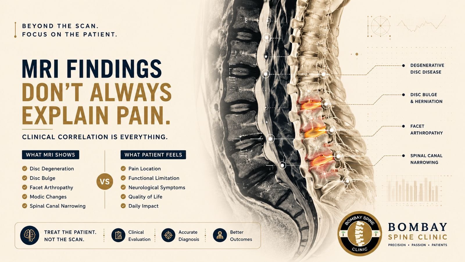
The Myth
A persistent misconception in spine care is that *lumbar MRI abnormalities directly correlate with pain, disability, or the need for intervention. Degenerative findings such as disc bulges, annular tears, Modic changes, or facet arthropathy are often interpreted as definitive pain generators—even in patients with minimal or no symptoms.
This imaging-first mindset has unintentionally fueled:
- Overdiagnosis
- Patient anxiety
- Unnecessary injections or surgery
- Delayed functional rehabilitation
The reality: MRI findings are extraordinarily common in asymptomatic individuals.
The Evidence
The landmark systematic review published in The Lancet and the widely cited Brinjikji et al. meta-analysis demonstrated that age-related degenerative MRI findings are prevalent even in pain-free populations.
Key findings included:
- Disc degeneration in over 90% of adults above 60
- Disc bulges in asymptomatic patients across nearly all age groups
- Annular fissures and facet degeneration frequently incidental
The implication is critical:
Structural abnormalities do not equal symptomatic pathology.
Guidelines from North American Spine Society and AO Spine consistently emphasize that imaging must be interpreted within the framework of:
- Clinical examination
- Neurological findings
- Functional impairment
- Symptom chronology
Yet the myth persists for several reasons:
- MRI Availability Has Outpaced Clinical Judgment
Advanced imaging is more accessible than ever, leading to premature MRI ordering before adequate clinical stratification.
- Defensive Medicine
Clinicians frequently fear missing serious pathology, even when red flags are absent.
- Patient Expectation
Many patients equate “finding something on MRI” with diagnostic certainty and expect imaging early in the treatment pathway.
- Incidental Findings Become Anchors
Once a degenerative change is identified, it can bias subsequent clinical decision-making—even when unrelated to symptoms.
The Modern Protocol
- Start With Symptom Pattern Recognition
Differentiate:
- Mechanical low back pain
- Radiculopathy
- Neurogenic claudication
- Inflammatory pain
- Red-flag pathology
Pain distribution and functional limitation remain more valuable than isolated imaging abnormalities.
- Screen for Red Flags Before Imaging
Immediate MRI should be reserved for:
- Progressive neurological deficit
- Suspected infection
- Malignancy
- Cauda equina syndrome
- Significant trauma
In uncomplicated low back pain, early MRI rarely improves outcomes.
- Correlate Imaging With Clinical Anatomy
A radiological abnormality matters only if it explains:
- Dermatomal symptoms
- Neurological deficit
- Concordant provocative findings
For example:
- A left paracentral L5-S1 disc prolapse without S1 symptoms may be incidental.
- Severe stenosis on MRI can exist in minimally symptomatic patients.
- Prioritize Functional Outcomes
Modern spine care focuses on:
- Walking tolerance
- Work capacity
- Sleep quality
- Neurological preservation
- Quality of life
Not MRI aesthetics.
- Use MRI as a Surgical Planning Tool—Not a Screening Tool
Imaging becomes truly valuable when:
- Conservative management fails
- Neurological compression correlates clinically
- Intervention is being considered
MRI should support a diagnosis—not create one.
Conclusion
The future of spine care lies in clinical-radiological balance, not radiological absolutism. Over-reliance on lumbar MRI findings risks converting normal aging into disease and exposes patients to unnecessary interventions.
Evidence-based spine practice demands a return to disciplined clinical correlation, functional assessment, and selective imaging utilization. The best outcomes occur when clinicians treat the patient—not the scan.
Dr. Siddharth Katkade | Bombay Spine Clinic


Comments are closed